Dr. Albert’s primary research focus is on applying HP helium-3 and HP xenon-129 MRI to image ventilation function in the airways and alveoli of the lungs. In addition, his group has been developing the following innovative imaging technologies: (1) HP xenon-129 functional MRI (xenon fMRI) of the brain; (2) HP xenon fMRI of stroke and traumatic brain injury; (3) HP xenon biosensor MR imaging of breast tumours; (4) HP xenon biosensor MRI for early detection of lung cancer; and (5) HP xenon biosensor MRI for early detection of Alzheimer’s disease.
Our Research
RESEARCH OVERVIEW
Lung Imaging
Since the original invention by Dr. Albert and Colleagues in 1994, Hyperpolarized (HP) noble gas MRI was used for functional imaging of the lungs over the globe. Imaging data acquired using HP noble gas MRI can be used to calculated as local as global pulmonological parameters of the human lungs which can be used for diagnostics and treatment monitoring of lung diseases such as asthma, chronic obstructive pulmonary disease (COPD), and idiopathic pulmonary fibrosis (IPF). Figure 1 shows an example of HP 129Xe MR images from our laboratory obtained from the healthy volunteer (A) and patients with asthma (B), COPD (C,D), and asbestosis (E). The ventilation defects associated with the disease progression can be clearly observed (B-E).
Another approach how to conduct functional MRI imaging of human lungs relies on the use of inert fluorinated gases. These gases are safe for inhalation and can be mixed with oxygen in any proportions. In addition, inert fluorinated gases are significantly cheaper compared to noble gases and require no polarization prior to inhalation. Although the fluorine signal is significantly smaller compared to HP noble gas signal, the quality of 19F images is sufficient for diagnostics purposes. To improve the performance of inert fluorinated gas MRI the study of gases with high number of fluorine atoms is needed as well as optimization of imaging parameters.

Figure 1. Examples of hyperpolarized xenon-129 lung images acquired in our lab
HP noble gas MRI and inert fluorinated gas MRI uses no ionizing radiation and it does not require exposing patients to the risks of chemical contrast agents that are sometimes used with conventional MRI. This allows HP gas and inert fluorinated gas MRI to be used to image patients repeatedly over time, allowing physicians to monitor how medical conditions progress, and to assess the effectiveness of specific treatments. We pursue our investigations on HP and inert gas MRI in collaborations with physicians, and with biomedical scientists, engineers, chemists, and physicists at TBRRI and a number of other universities and corporations. Currently, our investigations include work in the following areas:
- Optimization of imaging parameters for HP noble gas and inert fluorinated gas MRI.
- 19F lung MRI using octafluorocyclobutane development.
- Development of HP 129Xe imaging guidance for radiation therapy to minimize the radiation induced lung injury.
- Image performance comparison of the inert fluorinated gases.
Biosensors
Molecular imaging is a medical imaging field that has a potential to revolutionize the diagnosis and management of the disease. Molecular imaging is based on the injection of the imaging biosensor (molecular probe) which selectively binds with high affinity to a disease site and quickly removes from the rest of the body. Therefore, the signal originated from the probe perfectly correlated with a pathology location. Traditionally, MRI was not suitable for molecular imaging due to the poor sensitivity of this medical imaging modality.
Recently, the novel approach of molecular imaging using hyperpolarized (HP) xenon-129 (129Xe) chemical exchange saturation transfer (HyperCEST) effect. This methodology requires biosensors conjugated with supramolecular macrocycle (cage molecule) which can effectively encapsulate HP 129Xe. Due to constant exchange between the cage and the dissolved pool, it becomes possible to produce sufficient contrast by irradiating the encapsulated 129Xe using a narrow bandwidth radiofrequency pulse. The signal enhancement acquired using HyperCEST sufficient for molecular imaging purposes and recently we did the first HyperCEST imaging in the living rat (Figure 1). Together with Dr. DeBoef’s group from the University of Rhode Island we developing HyperCEST biosensors based on cucurbit[6]uril, gamma-cyclodextrin, and pillar[5]arene supramolecular cages. Figure 2 illustrated the structure of the macromolecules used in our studies.
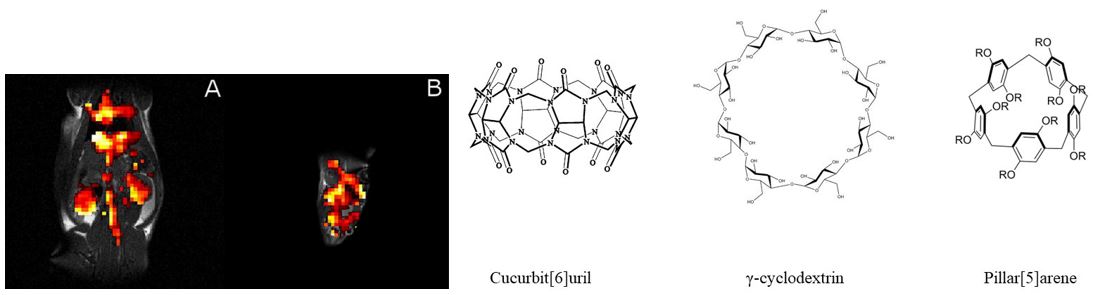
Figure 2. The hyperCEST image of the rat abdomen (A) and brain (B) acquired using Cucurbit[6]uril molecule. The chemical structures of supramolecular cages used for HyperCEST effect detection in our lab are shown on the right.
We are currently working to achieve the following goals:
- To develop a functionalized HyperCEST biosensor for detection of amyloid-beta deposition in the brain.
- To develop a functionalized HyperCEST biosensor for detection of hyperphosphorylated Tau protein in the brain.
- To develop fluorinated biosensors suitable for Alzheimer’s disease detection in the living organism.
Brain Imaging
The Albert group is intensively working on the HP 129Xe brain and cerebral blood flow imaging investigation. Due to the ability of HP 129Xe to dissolve in blood and travel to a highly perfused organs, the HP 129Xe can be potentially used as a contrast agent for quantitative blood flow measurement and brain imaging. The main goal of this project is to develop a method that is capable to measure a cerebral activity and cerebral perfusion directly with a high sensitivity and high contrast.
Albert and colleagues applied HP 129Xe MRI to image ischemic areas of the brain following middle cerebral artery occlusion (Zhou et. al. 2011). Following this work, the first use of HP 129Xe functional MRI was demonstrated in living rats. The activated areas of the rat brain were observed as a response to the pain stimulation (Mazzanti et. al. 2011). Recently, Albert’s group was able to study the perfusion changes associated with Alzheimer’s disease using HP 129Xe spectroscopy (Figure 3) and imaging (Figure 4).

Figure 3. (A) Stack plot of dynamic 129Xe NMR spectra for healthy controls (blue) and AD patients (red). (B) Topographic “streak” plot of (A) depicting the NMR dynamic spectra from the top with SNR in “hotter” colors. Notice a higher SNR in AD patients for a longer time than that of the healthy controls. SNR of 129Xe-WM (C) and 129Xe-GM (D) as a function of time for healthy controls (blue) and AD participants (red). The participants inhaled 500 mL of HP 129Xe and held their breath for 20 s. 129Xe MRS from the brain region was acquired every 2 s. Notice an increase in 129Xe signal after approximately 10 s as the 129Xe reached the brain. At 20 s, the participant exhaled and the 129Xe signal began to decrease at different rates for AD participants vs. healthy controls for WM and GM. The image was published in Diagnostics 2018,8, 41. doi:10.3390/diagnostics8020041
It was demonstrated for the first time that dynamic HP 129Xe imaging can quantitively measure the cerebral perfusion difference between the healthy brains and Alzheimer disease brains (Hane et. al. 2018).

Figure 4. Axial and sagittal 129Xe MRI of healthy controls and AD participants. An observably higher SNR was obtained for healthy controls relative to AD participants. The image was published in Diagnostics 2018, 8, 41. doi:10.3390/diagnostics8020041